Introducing HURT! App: Free After Hours Virtual Orthopedic Consultations

When you’re injured, the last thing you want is to wait days or weeks for advice. Orthopedic virtual consultations via the HURT! app provide a timely solution, offering professional care from the comfort of your home or wherever the injury happens. In this blog, we’ll explain virtual consultations, discuss when to seek one and introduce HURT!, a new app designed for instant orthopedic advice.

What is a Virtual Orthopedic Consultation?

Having a best friend, neighbor, or family member who works in the medical field brings peace of mind. If an accident happens, you can ask them for advice anytime, free of charge. But not everyone has that option, and even if you do, sometimes that person can only do so much.

That’s where virtual orthopedic consultations come in. A virtual orthopedic consultation is like having a doctor friend available 24/7.

Through virtual visits on the HURT! App, you can get immediate, professional advice on orthopedic concerns. The specialist can guide you on whether you need to visit the emergency room or see a doctor. It’s completely free, with no insurance billing involved. Plus, if you need an appointment, the specialist will facilitate an appointment by having the practice contact you directly.

When should you seek an Orthopedic Virtual Visit?

Virtual orthopedic consultations are ideal for non-emergency situations requiring professional advice. A virtual consultation can provide quick guidance on the next steps if you experience a sudden injury that isn’t life-threatening but still requires immediate attention. Additionally, if an injury occurs after regular office hours and you’re unsure whether to visit the emergency room or wait until morning, the HURT! app offers immediate professional advice on the appropriate course of action.

4 Benefits of Virtual Orthopedic Consultations

Virtual orthopedic consultations offer many advantages, transforming how patients access care. Here’s a closer look at the benefits you can expect:

  1. Cost-Effective: Virtual consultations, particularly through the HURT! app, are completely free of charge, and do not involve insurance billing. This can save you money, especially if it helps avoid a costly ER visit when it’s not necessary. By receiving professional advice at no cost, you can better manage your healthcare expenses.
  2. Saves Time: One of the greatest benefits of virtual consultations is the time saved. Instead of waiting hours at the ER or urgent care clinic, you can get professional guidance instantly. The HURT! app is available 24/7, providing a quick way to get a professional opinion whenever you need it without the wait.
  3. Convenient: The convenience of virtual consultations cannot be overstated. You can use the HURT! app anywhere and anytime, whether you’re at home, at work, or on the go. Additionally, you can access HURT! on a desktop through a web browser, making getting the care you need from any device easy.
  4. Peace of Mind: Having immediate access to professional advice brings peace of mind. With the HURT! app, you can feel confident about where to get care if needed. Knowing you have expert guidance available anytime helps reduce anxiety and ensures you make informed health decisions.

How much does a Virtual Orthopedic Consultation Cost?

The cost of a virtual orthopedic consultation is completely free when using the HURT! App. No charges or insurance billing are involved, allowing you to receive professional advice without any financial burden.

Introducing the HURT! App for Quick, Free Orthopedic Advice

Accidents and injuries don’t follow a schedule, often occurring when it’s least convenient. Waiting for an appointment with a specialist or spending hours in the ER can be grueling when you’re in pain. Thankfully, there is another option.
TSAOG Orthopedics and Spine partnered with the HURT! app to offer FREE, virtual after-hours access to orthopedic specialists. We’re here 24/7, 365 days a year, and are proud to connect you with the right orthopedic care when needed.

The HURT! app lets users connect with an orthopedic expert in minutes. The app makes access quick and easy, ensuring you receive immediate guidance without the need to leave your home. Whether it’s a sprain, strain, or any other orthopedic concern, the app provides professional advice and navigation to schedule an appointment with one of our orthopedic specialists if necessary.

Key Features of the HURT! App:

  • Free Access: The app is free to download and use, keeping your money in your pocket while receiving top-notch orthopedic advice.
  • User-Friendly Interface: Easily navigate the app to connect with experts quickly and efficiently.
  • Quick Response: Get timely responses to your queries, ensuring you receive the care you need without delay.
  • 24/7 Availability: Access to orthopedic specialists is available 24/7, every day of the year, including holidays.
  • Avoid Unnecessary ER Visits: Receive immediate guidance to avoid possibly unnecessary visits to the emergency room, saving time and resources.

Don’t wait for an injury to happen. Download HURT! Now for free, fast, virtual access to orthopedic specialists.

What is a Ganglion Cyst: Causes and Treatment Options

Reviewed by Doctor David Espinosa

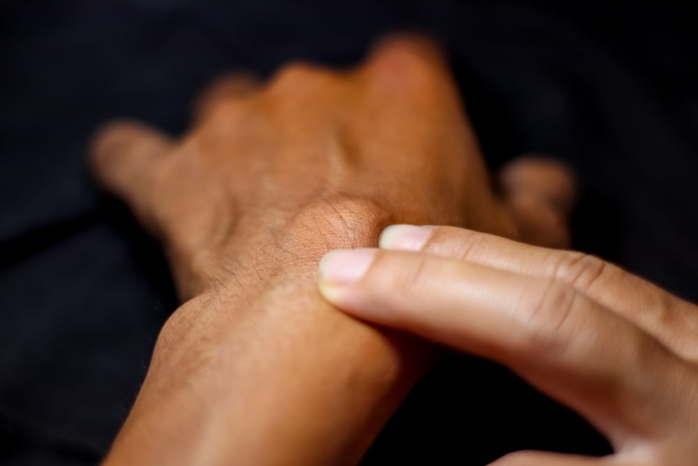
Ganglion cysts are noncancerous lumps that commonly develop along the tendons or joints of the wrists or hands. They can also appear on the ankles and feet. These cysts are filled with a jelly-like fluid and can vary in size. Understanding ganglion cysts, their causes, symptoms, and available treatments can help manage this condition effectively. Join us as we explain the causes, treatment options, and when to see a doctor.

What is a ganglion cyst?

A ganglion cyst is a round, fluid-filled lump typically forming near the joints or tendons. These cysts are usually noncancerous and range in size from a pea to a golf ball. The fluid inside a ganglion cyst is thick and sticky, similar to the fluid that lubricates joints and tendons.

Ganglion cysts form when synovial fluid leaks out of a joint or tendon sheath and accumulates in a sac. The exact mechanism is not fully understood, but it is believed that trauma or repetitive stress on a joint may contribute to the formation of these cysts. The cysts can appear suddenly or develop gradually over time.

Common Locations

  • Wrist and Hand: The most common location for ganglion cysts on the wrist or hand. They develop on the back of the wrist or the palm side.
  • Ankle and Foot: Less commonly, ganglion cysts can form on the top of the foot or around the ankle.
  • Other Areas: Although rare, ganglion cysts can also appear on the knee, shoulder, or spine.

Ganglion Cyst Causes

The exact cause of ganglion cysts is unknown, but several factors may contribute to their development.

Trauma

One theory is that trauma causes the joint tissue to break down, forming small cysts that then join together into a larger, more noticeable mass. Repeated injuries or overuse of a particular joint can increase the risk of developing a ganglion cyst. Activities like heavy lifting or contact sports can exacerbate this risk.

Joint or Tendon Irritation

Chronic irritation of a joint or tendon can lead to the formation of a ganglion cyst. The irritation can be due to repetitive motion, such as typing or certain sports activities. The constant stress and movement cause the joint to react by producing excess synovial fluid, forming the cyst.

Genetic Factors

Ganglion cysts may have a genetic component. If a family member has had one, there is an increased likelihood that others in the family will develop one as well.

Underlying Joint or Tendon Issues

Conditions that affect the joints or tendons, such as arthritis, can increase the risk of developing ganglion cysts. These conditions cause changes in the joint or tendon environment, which may promote cyst formation. Chronic joint inflammation creates an environment conducive to cyst development.

Ganglion Cyst Symptoms

Ganglion cysts can cause various symptoms, depending on their size and location.

Visible Lump

The most noticeable symptom of a ganglion cyst is a visible lump. This lump is usually round or oval, and its size can fluctuate. The cyst may grow larger with increased activity and shrink when at rest. The lump can be soft or firm to the touch.

Pain and Discomfort

While ganglion cysts are often painless, they can cause pain or discomfort if they press on nerves. This pain can be sharp or dull and may increase with joint movement or pressure on the cyst. Activities that involve repetitive motion or strain can exacerbate the pain.

Reduced Range of Motion

A large cyst or one located near a joint can limit the range of motion, making it difficult to move the affected joint fully. This can interfere with daily activities and tasks, making simple movements like gripping objects or bending the wrist challenging.

Numbness and Tingling

If a ganglion cyst presses on a nerve, it can cause numbness, tingling, or muscle weakness in the affected area. These symptoms are more common when the cyst is located near the wrist or hand. Persistent numbness and tingling can significantly impact hand function.

Ganglion Cyst Treatment Options

Treatment for ganglion cysts varies based on the severity of symptoms and the patient’s preferences. Both non-surgical and surgical options are available.

Non-Surgical Treatments

  • Observation

Often, ganglion cysts do not require treatment and may resolve independently. If the cyst is not causing pain or interfering with daily activities, a doctor may recommend a period of observation to monitor any changes.

  • Immobilization

A brace or splint can help immobilize the affected joint, reducing activity and potentially shrinking the cyst. Immobilization can also help alleviate pain by minimizing joint movement.

  • Aspiration

Aspiration uses a needle to drain the fluid from the cyst. The procedure is typically performed in a doctor’s office under local anesthesia. While aspiration can provide relief, the cyst may recur if the underlying cause is not addressed.

Surgical Treatments

  • Ganglionectomy

A ganglionectomy is a surgical procedure to remove the cyst. This option is considered when non-surgical treatments are ineffective or the cyst causes significant pain or limitations. Depending on the cyst’s location and size, the surgery can be performed using an open technique or arthroscopically.

  • Risks and Recovery

As with any surgery, there are risks associated with ganglionectomy, including infection, nerve damage, and scarring. Recovery time varies, but most patients can return to normal activities within a few weeks, with full recovery taking a few months.

When To Seek a Doctor for Ganglion Cyst

It is important to seek medical advice if you experience:

  • Persistent Pain: Pain that does not improve with over-the-counter pain relievers or home treatments should be evaluated by a doctor to determine the underlying cause and appropriate management.
  • Changes in Size or Shape: If the cyst grows rapidly or changes in shape, it should be examined to rule out other conditions and ensure it is not indicative of a more serious issue.
  • Numbness or Tingling: Any numbness, tingling, or weakness in the affected area warrants a medical evaluation to assess potential nerve involvement and prevent further complications.
  • Interference with Daily Activities: If the cyst limits your ability to perform daily tasks or participate in activities, medical intervention may be necessary to restore function and improve your quality of life.

Common Ganglion Cyst FAQ’s

Ganglion cysts can be a source of concern and curiosity for many people. To help you better understand the condition, we have compiled answers to some of the most frequently asked questions about ganglion cysts. Whether you’re dealing with a ganglion cyst yourself or seeking information for a loved one, these answers will help address common concerns and provide useful insights into managing this condition.

What happens if a ganglion cyst is left untreated?

In many cases, ganglion cysts are harmless and do not require treatment. They may remain the same size, grow, or even disappear on their own. However, if a cyst causes pain, limits movement, or affects daily activities, it should be evaluated by a healthcare professional. Leaving a symptomatic cyst untreated can lead to chronic discomfort and potential complications.

What shrinks a ganglion cyst?

Ganglion cysts may shrink or disappear with rest and reduced activity of the affected joint. Immobilization with a brace or splint can also help. Aspiration, where the cyst is drained of its fluid, can temporarily shrink the cyst, but there is a chance it may recur. In some cases, the cyst may resolve without any intervention.

What worsens ganglion cyst?

Activities that put stress on the affected joint can worsen a ganglion cyst. Repetitive motions, such as typing, sports, or heavy lifting, can increase the size of the cyst or exacerbate symptoms. Trauma or injury to the area can also worsen the cyst. It is important to modify activities and protect the joint to prevent aggravation.

How long do ganglion cysts last?

The duration of a ganglion cyst varies. Some cysts may resolve within a few weeks to months without treatment, while others may persist for years, especially if they are not causing significant symptoms. Treatment can speed up the resolution, but recurrence is possible. Regular monitoring and follow-up with a healthcare provider can help manage the condition.

Do ganglion cysts go away by themselves?

Ganglion cysts can go away on their own, especially if they are small and do not cause symptoms. Spontaneous resolution is more common in cysts that develop without a clear cause. However, cysts that are large, painful, or associated with joint or tendon issues are less likely to disappear without treatment. Observation and conservative management are often recommended for asymptomatic cysts.

By recognizing the signs and symptoms of ganglion cysts and seeking appropriate treatment, individuals can reduce pain, restore function, and prevent recurrence.

If you have any concerns about a lump or cyst near your joints, schedule an appointment with TSAOG today.

What is Shoulder Instability?

Reviewed by Dr. Robert Hartzler Robert U. Hartzler, M.D., M.S. – TSAOG Orthopaedics & Spine.

Key Takeaways:

  • “Shoulder instability” means that the ball of the shoulder joint is not staying properly in the socket
  • The shoulder commonly experiences instability because it is a “loose joint” that is highly mobile.
  • Shoulder instability is a complex problem: virtually every aspect of its management is debated amongst shoulder experts
  • Any patient, regardless of age, sex, or activity level, who has had a serious shoulder instability event (ball coming out of the socket) should seek care with an orthopaedic surgeon who can provide guidance about management (imaging and treatment options)
  • Shoulder instability is not a “DIY” condition, since incorrect management can result in further damage to the shoulder and potentially risk worsening of the condition
  • Surgery is often needed to prevent further instability events, especially for young athletes
  • Non-surgical management can require the care of specialized therapists and/or therapy devices such as e-stim
  • For athletes desiring to return to high-risk collision sports (e.g. football, rugby, ice hocky) return to play testing can lower the rate of recurrent instability

Shoulder instability happens when the structures around the shoulder joint do not work to keep the ball within its socket. Instability occurs on a spectrum from dislocation (ball completely out of socket), to subluxation (ball partly out of socket), to apprehension (feeling that the ball might come out of socket). The typical instability patient doesn’t feel pain except when instability is happening; however, chronic instability can lead to painful damage of the bones or soft tissues around the joint.

Understanding the nature of this condition is crucial for managing and treating it effectively. In short, shoulder instability is a complex problem: virtually every aspect of its management is debated amongst shoulder experts. This includes the terminology of shoulder instability, the meaning of physical examination findings, the correct imaging studies to obtain in its diagnosis, the right treatment options, and the best pathway to return to sports for athletes.

Any patient, regardless of age, sex, or activity level, who has had a serious shoulder instability event (ball coming out of the socket) should seek care with an orthopaedic surgeon who can provide guidance about management (imaging and treatment options).

Join us as we explore the topic of shoulder instability: anatomy of the shoulder, symptoms, causes, treatment options, and when to see a doctor.

Shoulder Anatomy

Before we dive into shoulder instability, let’s briefly discuss shoulder anatomy.

The shoulder joint, or the glenohumeral joint, consists of three main bones:

  1. humerus (or upper arm bone)
  2. scapula (or shoulder blade)
  3. clavicle (or collarbone.)

The head of the humerus fits into a shallow socket in the scapula referred to as the glenoid. This socket is surrounded by a ring of cartilage called the labrum, which helps deepen the socket and stabilize the joint.

Ligaments, which are strong bands of tissue, connect the bones and provide stability by limiting the joint’s movement at the end ranges of motion. A group of muscles and tendons known as the rotator cuff surrounds the shoulder joint and provides support and movement when the joint is not at the end ranges. When any of the soft tissues (labrum, ligaments, or muscle-tendons units) holding the shoulder in place become damaged or weakened, the joint can become unstable.

What is chronic shoulder instability?

Chronic shoulder instability is a condition in which the shoulder repeatedly slips out of place. This instability can be due to trauma or congenital issues. When the shoulder is chronically unstable, it can be progressively damaged such that each instability event makes another one more likely to happen.

Types of Shoulder Instability

Understanding the different types of shoulder instability can help diagnose and determine an appropriate treatment plan.

Traumatic Shoulder Instability

This type occurs when a sudden injury or trauma, such as a fall or sports injury, causes the shoulder to dislocate. Traumatic shoulder instability is often associated with high-impact activities and is common in contact sports like football, rugby, and wrestling.

A dislocation occurs when the head of the humerus is forced out of the glenoid socket. Dislocation can happen in various directions: anterior (forward), posterior (backward), or inferior (downward). Anterior dislocations are the most common, accounting for the vast majority of all shoulder dislocations. Anterior dislocation usually happens when the arm is forced into an awkward position, such as during a fall on an outstretched hand.

Consider a football player who experiences a dislocation during a tackle. The impact forces the shoulder out of its socket, tearing the ligaments and potentially causing a labral tear. Even after initial treatment, the shoulder remains vulnerable to future dislocations, especially if the player returns to the sport without adequate rehabilitation.

Atraumatic Shoulder Instability

This instability happens without a significant injury and can develop gradually due to repetitive overhead activities or inherent looseness in the shoulder joint. It is often seen in individuals who engage in activities that involve repetitive motion, such as swimming, gymnastics, or volleyball.

For example, a competitive swimmer may develop atraumatic instability due to the repetitive overhead motion of the swimming strokes. Over time, the shoulder ligaments become stretched, leading to a feeling of looseness and pain during activity. The elongation can progress to chronic instability without proper intervention, impacting the athlete’s performance and quality of life.

Acquired Shoulder Instability

This type results from repetitive movements, often seen in athletes who participate in activities that require frequent overhead motions, leading to gradual stretching of the shoulder ligaments. Acquired instability is often a combination of both traumatic and atraumatic factors.

Take gymnasts, for instance, who frequently perform routines that require significant shoulder strength and flexibility. The constant strain on the shoulder joint can lead to acquired instability, where the shoulder becomes loose over time.

Common Symptoms of Shoulder Instability

Recognizing the symptoms of shoulder instability is crucial for early intervention and treatment.

  • Frequent Dislocations: Repeated dislocations are a hallmark of shoulder instability. These can occur during physical activity or simple movements, such as reaching overhead or lifting objects.
  • Sensation of Looseness: Many individuals with shoulder instability report a feeling that their shoulder is loose or about to give way. This sensation can be unsettling and affect daily activities.
  • Pain: Pain can range from a dull ache to sharp, intense pain, especially during or after physical activity. Chronic pain is common in cases of long-standing instability.
  • Weakness: Muscle weakness, particularly in the shoulder and upper arm, can result from recurrent dislocations and the associated damage to the stabilizing structures.
  • Limited Range of Motion: Another result of chronic instability is difficulty moving the shoulder through its full range of motion. The limitations can be due to pain, muscle guarding, or mechanical blockages from damaged tissue.

Common Causes of Shoulder Instability

Understanding the underlying causes of shoulder instability can help develop effective prevention and treatment strategies.

Previous Dislocations

Prior shoulder dislocations can weaken the ligaments, making the joint more prone to future dislocations. Each dislocation stretches the ligament further, reducing its ability to stabilize the joint.

Repetitive Strain

Activities that involve repetitive overhead motions, such as swimming or throwing sports, can stretch the shoulder ligaments over time. This repeated strain can lead to gradual loosening of the joint, increasing the risk of instability.

Congenital Factors

Some individuals are born with naturally loose ligaments, making their shoulders more prone to instability. This condition, known as ligamentous laxity, can contribute to atraumatic and acquired shoulder instability.

Trauma

A sudden injury, such as a fall or a blow to the shoulder, can cause the shoulder to dislocate and become unstable. Traumatic events often damage the ligaments and other stabilizing structures, leading to long-term instability.

Shoulder Instability Treatment Options

Treatment for shoulder instability varies based on the severity of the condition, the patient’s activity level, and the underlying cause. Both non-surgical and surgical options are available.

Non-Surgical Treatments

Physical Therapy

Physical therapy is a cornerstone of non-surgical treatment for initial treatment of shoulder instability for certain patients with at low risk for recurrent instability. Alternatively, therapy and return to play once symptom free can be a poor choice for high risk patients for whom surgery should be strongly considered as initial treatment to prevent recurrence.

A structured rehabilitation program focuses on strengthening the shoulder muscles, improving joint stability, and restoring the range of motion.

  • Strengthening Exercises: Specific exercises target the rotator cuff and scapular muscles to enhance stability. Examples include external rotations, internal rotations, and scapular retractions.
  • Flexibility Exercises: Stretching exercises help maintain and improve the range of motion. These can include doorway stretches and cross-body shoulder stretches.
  • Proprioception Training: Exercises that enhance the body’s ability to sense the position of the shoulder joint can reduce the risk of dislocation. Balance exercises on unstable surfaces, such as a Bosu ball, are commonly used.

Medication

Anti-inflammatory medications can help reduce pain and swelling, making it easier for patients to participate in physical therapy and daily activities. Commonly prescribed medications include nonsteroidal anti-inflammatory drugs (NSAIDs) like ibuprofen and naproxen.

Bracing

Wearing a shoulder brace can provide extra support and stability to the joint during physical activities. Braces are controversial in their ability to affect recurrence. They may benefit athletes psychologically who wish to continue their sport while managing shoulder instability.

Surgical Treatments

Surgical intervention is often necessary to restore stability to the shoulder joint or to repair damaged structures.

Arthroscopic Surgery

This minimally invasive procedure involves repairing or tightening the ligaments using small incisions. Arthroscopic surgery is often used to address labral tears and to reattach ligaments to the bone.

  • Bankart Repair: This procedure involves reattaching the torn labrum to the glenoid socket. It is commonly performed in patients with anterior shoulder instability.
  • Capsular Shift: This surgery tightens the overstretched capsule by folding and suturing the excess tissue, reducing the joint’s looseness.
  • Arthroscopic bone reconstruction: advanced techniques for bone reconstruction of the socket and humeral head are now available depending on the experience level of the surgeon with these techniques

Open Surgery

In more severe cases, open surgery might be required to reposition or reconstruct the shoulder structures. Open surgery allows for more extensive reconstructions and is often used for complex cases.

  • Latajet Procedure: This procedure involves transferring a portion of the coracoid process, along with its attached muscles, to the front of the glenoid. This provides additional stability and prevents dislocation.
  • Humeral head bone reconstruction: using donated bone to reconstruct the impacted bone of the ball of the shoulder

When To Seek a Doctor About Shoulder Instability

Any patient, regardless of age, sex, or activity level, who has had a serious shoulder instability event (ball coming out of the socket) should seek care with an orthopaedic surgeon who can provide guidance about management (imaging and treatment options).

It is crucial to seek medical advice if you experience:

  • Recurrent Shoulder Dislocations: Frequent dislocations can indicate significant instability and should be evaluated by a healthcare professional.
  • Persistent Pain or Discomfort: Ongoing pain that affects daily activities or sleep warrants a medical evaluation to determine the cause and appropriate treatment.
  • Sensation of Looseness or Instability: Feeling that the shoulder is loose or unstable, especially during physical activities, should prompt a visit to the doctor.
  • Difficulty Performing Daily Activities: If shoulder issues interfere with work, sports, or daily tasks, seeking medical advice is essential.

Understanding shoulder instability and its implications is key to managing the condition effectively. By recognizing the symptoms, identifying the causes, and seeking appropriate treatment, individuals can achieve improved shoulder function and quality of life.

If you are experiencing shoulder instability, schedule an appointment with TSAOG today.